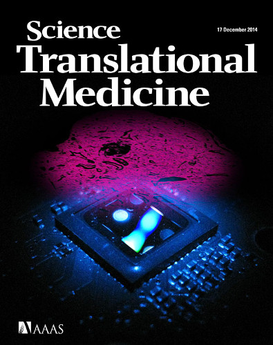
Wide-field Computational Imaging of Pathology Slides using Lensfree On-Chip Microscopy published in Science Translational Medicine (AAAS) (2014)
A. Greenbaum, Y. Zhang,

Optical examination of microscale features in pathology slides is one of the gold standards to diagnose disease. However, the use of conventional light microscopes is partially limited owing to their relatively high cost, bulkiness of lens-based optics, small field of view (FOV), and requirements for lateral scanning and three-dimensional (3D) focus adjustment. We illustrate the performance of a computational lens-free, holographic on-chip microscope that uses the transport-of-intensity equation, multi-height iterative phase retrieval, and rotational field transformations to perform wide-FOV imaging of pathology samples with comparable image quality to a traditional transmission lens-based microscope. The holographically reconstructed image can be digitally focused at any depth within the object FOV (after image capture) without the need for mechanical focus adjustment and is also digitally corrected for artifacts arising from uncontrolled tilting and height variations between the sample and sensor planes. Using this lens-free on-chip microscope, we successfully imaged invasive carcinoma cells within human breast sections, Papanicolaou smears revealing a high-grade squamous intraepithelial lesion, and sickle cell anemia blood smears over a FOV of 20.5 mm^2. The resulting wide-field lens-free images had sufficient image resolution and contrast for clinical evaluation, as demonstrated by a pathologist’s blinded diagnosis of breast cancer tissue samples, achieving an overall accuracy of ~99%. By providing high-resolution images of large-area pathology samples with 3D digital focus adjustment, lens-free on-chip microscopy can be useful in resource-limited and point-of-care settings.