Fluorescent Imaging of Single Nanoparticles and Viruses on a Smart-Phone published in ACS Nano (2013)
Q. Wei , H. Qi , W. Luo , D. Tseng ,
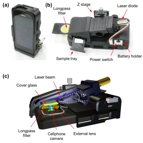
Optical imaging of nanoscale objects, whether it is based on scattering or fluorescence, is a challenging task due to reduced detection signal-to-noise ratio and contrast at subwavelength dimensions. Here, we report a field-portable fluorescence microscopy platform installed on a smart phone for imaging of individual nanoparticles as well as viruses using a lightweight and compact opto-mechanical attachment to the existing camera module of the cell phone. This hand-held fluorescent imaging device utilizes (i) a compact 450 nm laser diode that creates oblique excitation on the sample plane with an incidence angle of 75°, (ii) a long-pass thin-film interference filter to reject the scattered excitation light, (iii) an external lens creating 2× optical magnification, and (iv) a translation stage for focus adjustment. We tested the imaging performance of this smart-phone-enabled microscopy platform by detecting isolated 100 nm fluorescent particles as well as individual human cytomegaloviruses that are fluorescently labeled. The size of each detected nano-object on the cell phone platform was validated using scanning electron microscopy images of the same samples. This field-portable fluorescence microscopy attachment to the cell phone, weighing only 186 g, could be used for specific and sensitive imaging of subwavelength objects including various bacteria and viruses and, therefore, could provide a valuable platform for the practice of nanotechnology in field settings and for conducting viral load measurements and other biomedical tests even in remote and resource-limited environments.