High-throughput Imaging and Characterization of a Heterogeneous Cell Solution On a Chip published in Biotechnology and Bioengineering (2008)
By T-W. Su , S. Seo ,
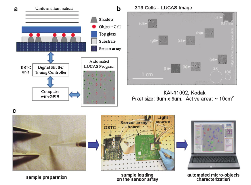
A high-throughput on-chip imaging platform that can rapidly monitor and characterize various cell types within a heterogeneous solution over a depth-of-field of 4 mm and a field-of-view of 10 cm2 is introduced. This powerful system can rapidly image/monitor multiple layers of cells, within a volume of 4 mL all in parallel without the need for any lenses, microscope-objectives or any mechanical scanning. In this high-throughput lensless imaging scheme, the classical diffraction pattern (i.e., the shadow) of each micro-particle within the entire sample volume is detected in less than a second using an opto-electronic sensor chip. The acquired shadow image is then digitally processed using a custom developed “decision algorithm” to enable both the identification of the particle location in 3D and the characterization of each micro-particle type within the sample volume. Throughexperimental results, we show that different cell types (e.g., red blood cells, fibroblasts, etc.) or other micro-particles all exhibit uniquely different hadow patterns and therefore can be rapidly identified without any ambiguity using the developed decision algorithm, enabling high-throughputcharacterization of a heterogeneous solution. This lensfree on chip cell imaging platform shows a significant promise especially for medical diagnostic applications relevant to global health problems, where compact and cost-effective diagnostic tools are urgently needed in resource limited settings.