Multi-color LUCAS: Lensfree on-chip cytometry using tunable monochromatic illumination and digital noise reduction published in Cellular and Molecular Bioengineering (2008)
By T-W. Su , S. Seo ,

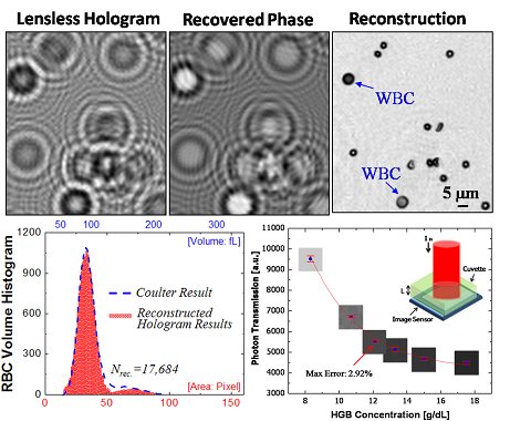
It has been recently demonstrated that rapid on-chip monitoring of a homogenouscell solution within an ultra-large field-of-field of ~10 cm2 isfeasible by recording the classical diffraction pattern of each cell in parallel onto an opto-electronic sensor array under incoherent white lightillumination. Here we present several major improvements over this previous approach. First, through experimental results, we illustrate that the use of narrowband short wavelength illumination (e.g., at ~300 nm) significantly improves the digital signal-to-noise ratio of the cell diffractionsignatures, which translates itself to a significant increase in the depth-of-field (e.g., ~5 mm) and hence the sample volume (e.g., 5 mL) that can be imaged. Second, we also illustrate that by varying the illumination wavelength, the texture of the recorded cell signatures can be tuned to enableautomated identification and characterization of a target cell type within a heterogeneous cell solution. Third, a hybrid imaging scheme that combines two different wavelengthsis also demonstrated to improve the uniformity and signal-to-noise ratio of the recorded cell diffraction images. Finally, we demonstrate that a further improvement in image quality can be achieved by utilizing adaptive digital filtering. These noteworthy improvements are especially quite important to achieve more reliable performance for rapid detection and counting of a target cell type among many other cells present within a heterogeneous sample volume of e.g., ~5 mL. This incoherent on-chip imaging platform may have a significant impact especially for medical diagnostic applications related to global health problems such as HIV monitoring.